
Flow Chart of Radiation Therapy
Consultation
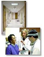 When you some to the clinic, a radiation oncologist will review your current problem, past medical history, past surgical history, family history, medications, allergies, lifestyle and an organ system by organ system audit making use of any relevant x-rays and lab tests that you already have had. He/she will also perform a physical examination that will then to assess the extent of your problem and judge your general physical condition to estimate any influence it may have over your response to radiation therapy.
Simulation
The simulation process will help the radiation team plan a course of treatment designed especially for you. The radiation oncologist uses the x-rays images and the information gathered during the simulation to design and individualise your treatment plan.
 Simulators are very precise machines that can duplicate the geometry of different types of treatment machines and by fluoroscopy and x-ray pictures allow us to visualise important structures inside patients. This information is translated into aiming points, distances and angles. With you lying on the simulator couch and with the use
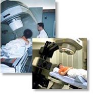 of light and laser projection systems we will place marks on your skin or on individually constructed immobilisation devices that will help level and position you in 3-dimensional space. Marking may be tattooed on the skin. Immobilisation devices will be constructed from one or more materials including thermally shaped plastics, vacuum forming beanbags, and plaster casts. You may need an appointment with the mould room for making of your immobilisation device.
Treatment Planning
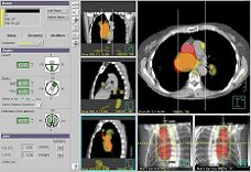
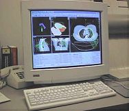
The treatment planning team includes the radiation oncologist, radiographer and physicist. Using all available information, including that obtained from simulation, the final plan will determine the type of radiation, its size and shape and the number of radiation beams, and how much radiation is given through each beam. Radiation beams must be shaped to match their intended targets with the collimator jaws. Depending on the desired shape, additional blocking of the beam may be required. The treatment planning computer then calculates the desired dose distribution taking into account the sparing of normal healthy tissue and the tolerance of the patient as a whole. The radiation oncologist approved the final plan and the treatment parameters are printed in a hardcopy with the electronic version sent to the treatment machine treatment record system via the hospital network.
 |
 |
Machine Quality Assurance
Treatment Routine
Monitoring Your Progress
Possible Side Effects
You are not radioactive and it is not harmful to anyone around you unless you have an implant of radioactive seeds, wire or ingested/infused radiopharmaceuticals. It is not unusual to feel tired during the time you are receiving radiation therapy. Many patients find it helpful to take an afternoon nap or get more sleep each night. Usually, you can continue your normal lifestyle. Your body is your best guide.